


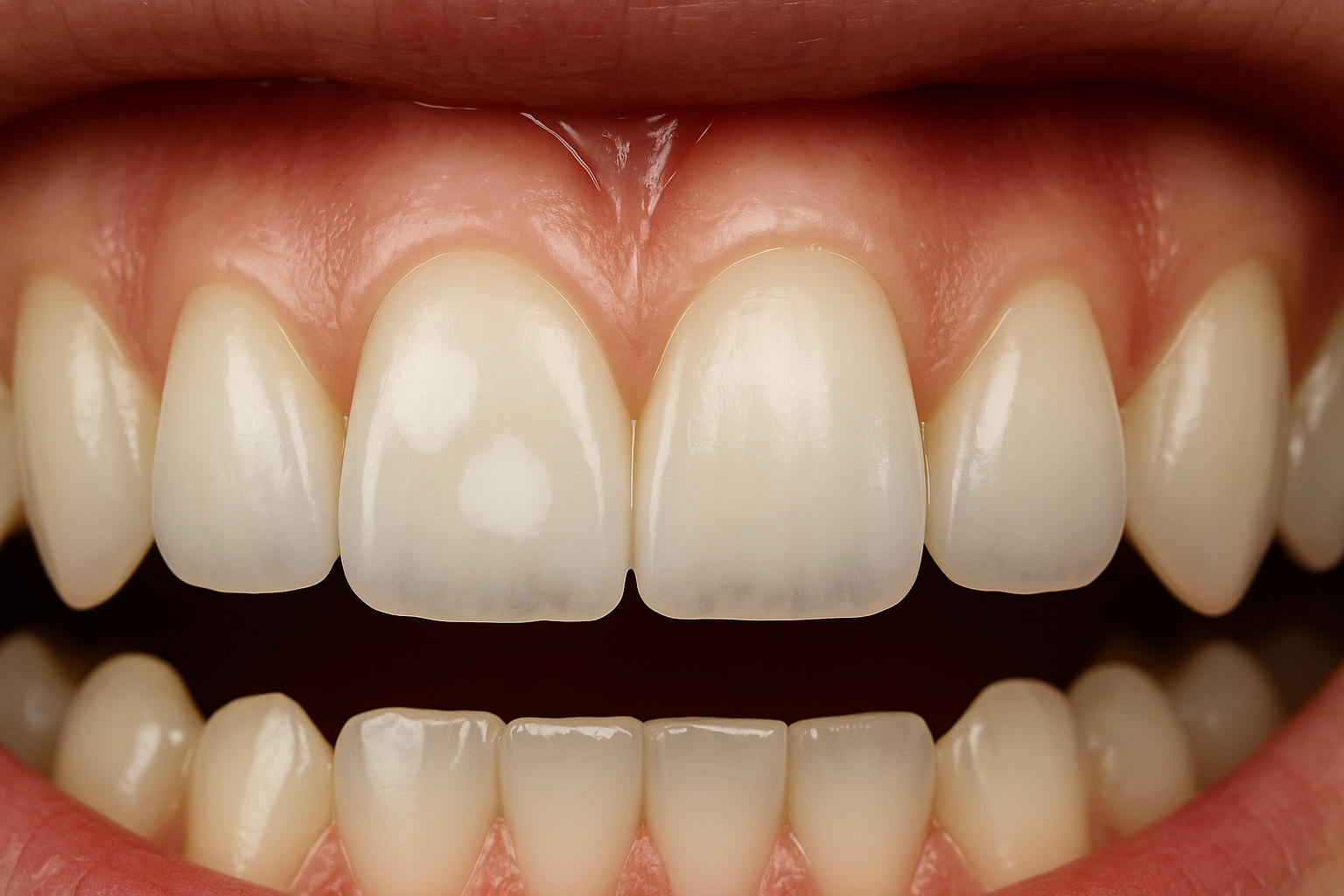
White spots on teeth are an aesthetic concern that increasingly worries patients of all ages. They may appear in both children and adults. While many people treat them merely as a cosmetic defect, in reality, they can be an early sign of developing disease, such as incipient caries. As a dentist with many years of experience, I know that proper diagnosis of such changes allows us to avoid more serious problems and plan effective treatment. In this article, I will explain: the main causes of white spots on teeth, how modern dental clinics diagnose them, which treatment methods bring the best results, and how to prevent the recurrence of this problem. Causes of White Spots on Teeth 1. Early Caries The most common cause of white spots is early-stage caries. At this stage, enamel demineralization occurs due to the action of bacteria and acids. Fortunately, the process can still be reversible if the patient visits the dentist promptly. 2. Fluorosis In children, excessive fluoride intake – from diet or supplements – may cause chalky white marks on tooth surfaces. This phenomenon is called fluorosis and requires dental assessment to determine appropriate treatment. 3. Demineralization after Orthodontic Treatment Patients who have worn fixed braces often notice white discolorations around brackets. These marks are areas of enamel demineralization, which can be treated effectively if detected early. 4. Enamel Developmental Disorders Congenital enamel defects (enamel hypoplasia) also manifest as white or yellowish spots. This is a developmental issue that requires an individualized approach. 5. Diet and Oral Hygiene Poor eating habits, frequent consumption of fizzy drinks, sweets, or acidic foods, as well as lack of professional dental cleaning, all increase the risk of white spots forming. Diagnosis of White Spots At Warsaw Dental Center, we use advanced diagnostic methods to precisely assess the nature of such changes: Microscope examination – allows us to observe enamel under magnification and assess the degree of demineralization, Intraoral photos and X-rays – help to check whether decay is developing beneath the enamel surface, Analysis of oral hygiene habits – helps determine the cause and tailor an effective treatment plan. It’s important to remember that the sooner a patient visits the dentist, the greater the chance to stop the progression and avoid drilling. Treatment of White Spots on Teeth 1. Enamel Remineralization In the earliest stages, we apply fluoride, calcium, and phosphate preparations to help rebuild the enamel structure. This treatment is completely painless. 2. ICON – Resin Infiltration This modern technique removes white spots without drilling. The dentist applies a special infiltrating resin that penetrates enamel micropores and restores natural tooth color. ICON is especially effective for spots after orthodontic treatment. 3. Teeth Whitening In some cases, controlled whitening helps even out the tooth shade and reduce the visibility of white spots. The procedure is performed under dental supervision to minimize sensitivity. 4. Porcelain Veneers If the changes are extensive or aesthetics require full correction, porcelain veneers are the best option. These thin ceramic shells provide teeth with perfect color and shape. Prevention : How to Avoid White Spots Regular professional cleaning at the dental office (at least once or twice a year). Daily brushing with fluoride toothpaste and flossing. Limiting sweets and acidic beverages in the diet. Routine dental check-ups – especially important for children and patients after orthodontic treatment. For pregnant women – regular check-ups to prevent demineralization and caries. Conclusion White spots on teeth may appear to be only a minor aesthetic issue, but they are often the first sign of caries or enamel mineralization disorders. Modern dentistry offers many solutions – from remineralization and ICON infiltration to aesthetic veneers. At Warsaw Dental Center, we approach every patient individually, using microscopes and advanced diagnostic tools to provide the best treatment. Urszula Leończak, MSc – Medical Director, Dentist

Wisdom teeth – what are they and how to deal with them?
Calculus deposits, also known as pulp stones, are calcifications that occur within the dental pulp, mainly in its chamber. Many of us are not aware of their presence until intense pain occurs. Therefore, we want to help in recognizing calculus deposits and effectively treating them. Where do calculus deposits come from? Calculus deposits, also known as dental pulp stones, are calcified deposits of mineral salts, which are a form of dental pulp degeneration. They most commonly occur in molars, lower incisors, and in teeth that are yet to erupt or are impacted. Calculus deposits typically develop in necrotic areas, mainly around the pulp chamber and root of the tooth. Although the exact causes of calculus deposits formation have not been fully elucidated, they are often associated with inflammatory conditions or tooth trauma. Calculus deposits can vary greatly in size, from microscopic changes invisible to the naked eye to large deposits that can lead to pulp chamber obliteration. Types of calculus deposits Due to differences in size, structure, and location, several types of calculus deposits are distinguished: Based on size and location: 1. Compact – located within the tooth chamber and visible on X-ray. 2. Dispersed – typically located in root canals and not visible on X-ray. Based on structure: 1. True – structurally similar to dentin, occurring relatively rarely. 2. False – lack a dentin-like structure, containing calcified cells that continuously expand due to increasing calcification. Based on location: 1. Loosely embedded – not attached to the tooth cavity walls and completely surrounded by pulp. 2. Wall-attached – partially connected to dentin. 3. Intrapulpal – completely surrounded by dentin. Symptoms of calculus deposits Calculus deposits often do not cause any pain symptoms for a long time, making them typically detected during routine dental visits or on X-rays. Only with the growth of calculus deposits, when they begin to compress the sensory nerve and blood vessels, does bothersome pain arise. Initially, this pain resembles discomfort associated with pulp or trigeminal nerve inflammation. As calculus deposits reach significant sizes, they can fill the entire tooth chamber, leading to pulp damage and necrosis. Confirmation of calculus deposits requires X-ray imaging. Treatment of calculus deposits Untreated and continuously developing calculus deposits can lead to serious health problems. In the case of small, painless calculus deposits, the dentist may recommend observation and regular check-ups. However, if pain symptoms arise, removal of the affected pulp and root canal treatment is recommended. This procedure is performed under anesthesia, making it painless. It is important to note that even small calculus deposits can complicate endodontic treatment, hindering access to the tooth chamber floor and canal openings. In such cases, removal may be necessary. Regular follow-up visits to the dentist are crucial as they allow for the prompt detection of any changes in the oral cavity. Finally, it is worth noting that calculus deposits are not associated with tumors. Many people mistakenly associate calculus deposits with benign tumor nodules due to the similarity in names to the term “odontoma.” However, despite growing within the pulp, calculus deposits are not neoplastic changes.
Panoramic photo of teeth – effective dental diagnostics
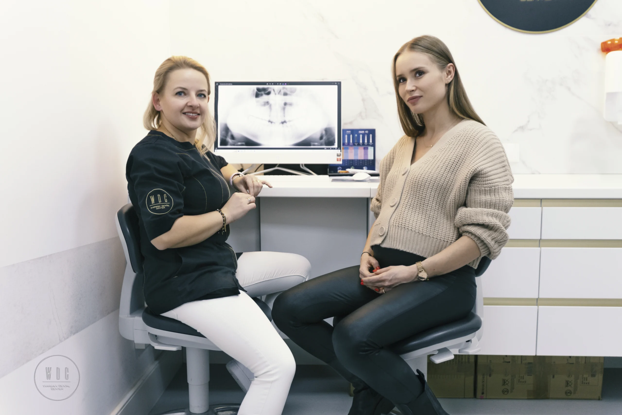
Dentist Aleksandra Kostrz discusses treatment with the patient about dental x-rays. Although an experienced dentist can detect many abnormalities using his eyes, he cannot see all the details. Therefore, to obtain greater certainty, it is recommended to perform a dental pantomography. This short and effective test allows you to detect even the smallest abnormalities, including the presence of cancer. Dental panoramic radiograph – for whom? A panoramic radiograph is a basic diagnostic test that is an important element of the patient’s medical documentation. Its aim is to quickly assess the condition of the teeth, periodontium, jaw bones, mandible, maxillary sinuses and temporal joints. They are performed using a device known as a pantomograph. This test detects diseases and problems invisible to the naked eye, such as: initial caries, impacted teeth, e.g. wisdom teeth, irregularities resulting from root canal treatment, cancer, cysts. A dental panoramic radiograph is also extremely useful in planning prosthetic treatment, such as dentures, crowns or bridges. Before starting orthodontic treatment, it is recommended to perform a panoramic radiograph, especially if the patient has unerupted wisdom teeth, which allows for appropriate preparation for their possible surgical removal. The course of the procedure of taking a panoramic X-ray The dental pantomography procedure takes several seconds and takes place in a separate room to prevent radiation from escaping. The patient is covered with a lead blanket, which, combined with a low dose of radiation, guarantees a high level of safety. No special preparations are required, such as the need to come on an empty stomach. Only movable elements, e.g. earrings, should be removed to avoid marks on the photo. The entire procedure is painless. During pantomography, the radiation-generating head moves simultaneously with the recorder, which allows obtaining a precise image of a specific part of the facial skeleton. The resulting image is then enlarged by approximately 15%.
Benefits of using a camera in dentistry
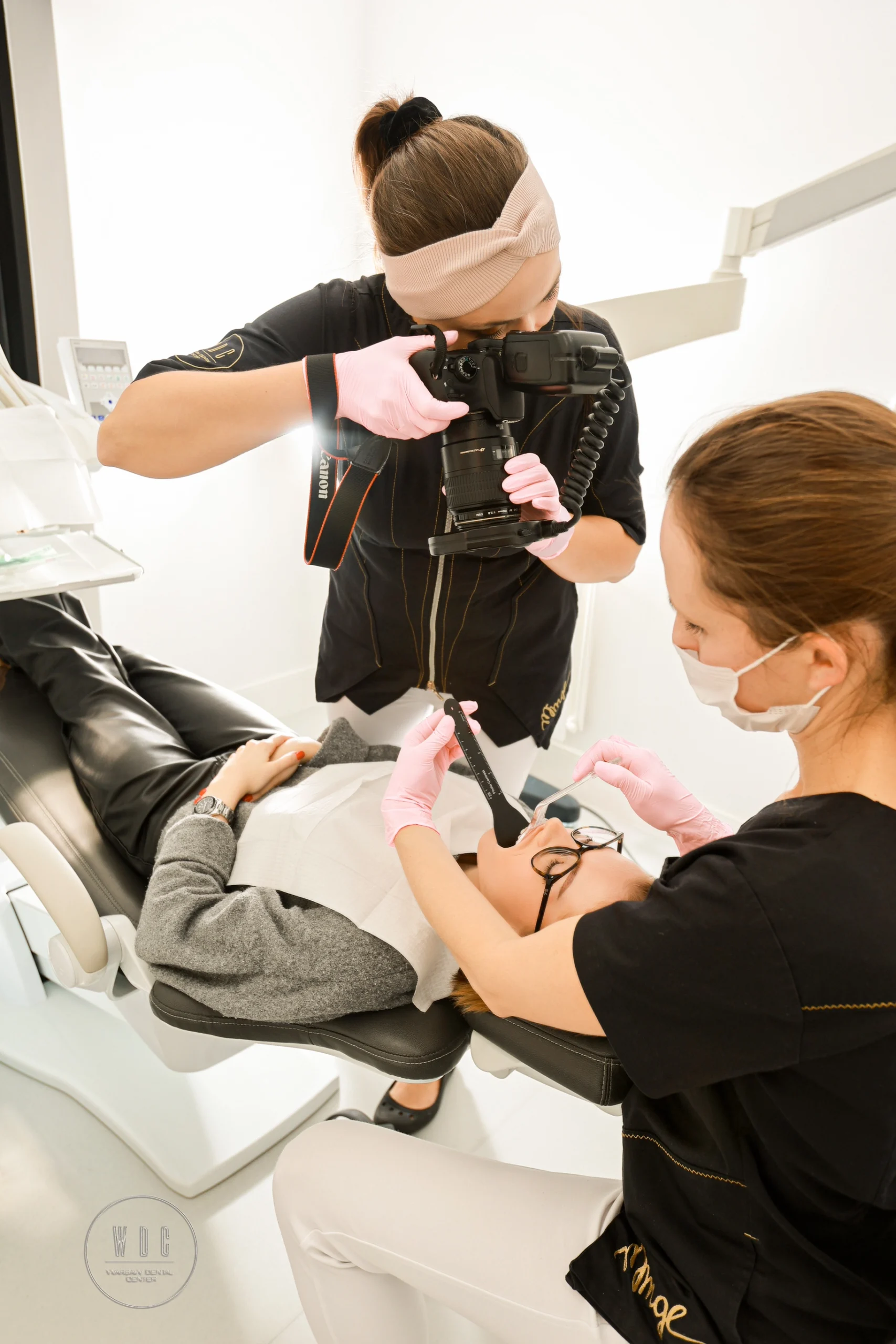
Dentist Natalia Rogulska takes a photo of the patient’s teeth Nowadays, cameras have become an indispensable tool in every modern dental clinic. Photographic documentation not only records the patient’s image, but opens the door to many benefits that can significantly improve the treatment process and communication with patients. Full photo protocol: The patient is always at your fingertips Taking a complete set of intraoral photos is a fundamental part of our practice. Thanks to this graphic document, we can return to the patient’s initial condition at any time. This is an invaluable value that allows us to closely monitor treatment progress and make more accurate therapeutic decisions. Collaboration and individual treatment plan Consultation with experts in various fields of dentistry is crucial to creating comprehensive and personalized treatment plans. Thanks to photographic documentation, we can collaborate more effectively, presenting case details and consulting with other specialists to determine the optimal therapeutic approach for the patient. Photos as an educational tool Photos can convey more than a thousand words. Photographs allow patients to better understand the current state of their oral health and the need for any changes. Often, what may be difficult to understand in conversation becomes clear and distinct in a photo. This is an excellent educational tool to help patients make more informed decisions about their treatment. Track your progress and be satisfied with your smile’s evolution Photos are also an excellent tool for assessing treatment progress. By comparing before and after photos, both doctor and patient can see the evolution and results of the therapy. Some smile transformations require time and systematic work, but the effects are extremely satisfying. Patients often forget what they looked like at the beginning of treatment, which makes the final visit an exciting and touching moment when they can compare their past and present smiles. The use of a camera in our dental practice is not only a documentation tool, but above all a communication and motivational tool for both us and our patients. Each photo is a step towards a more beautiful, healthier smile!
Dental microscope – more than just “special tasks”
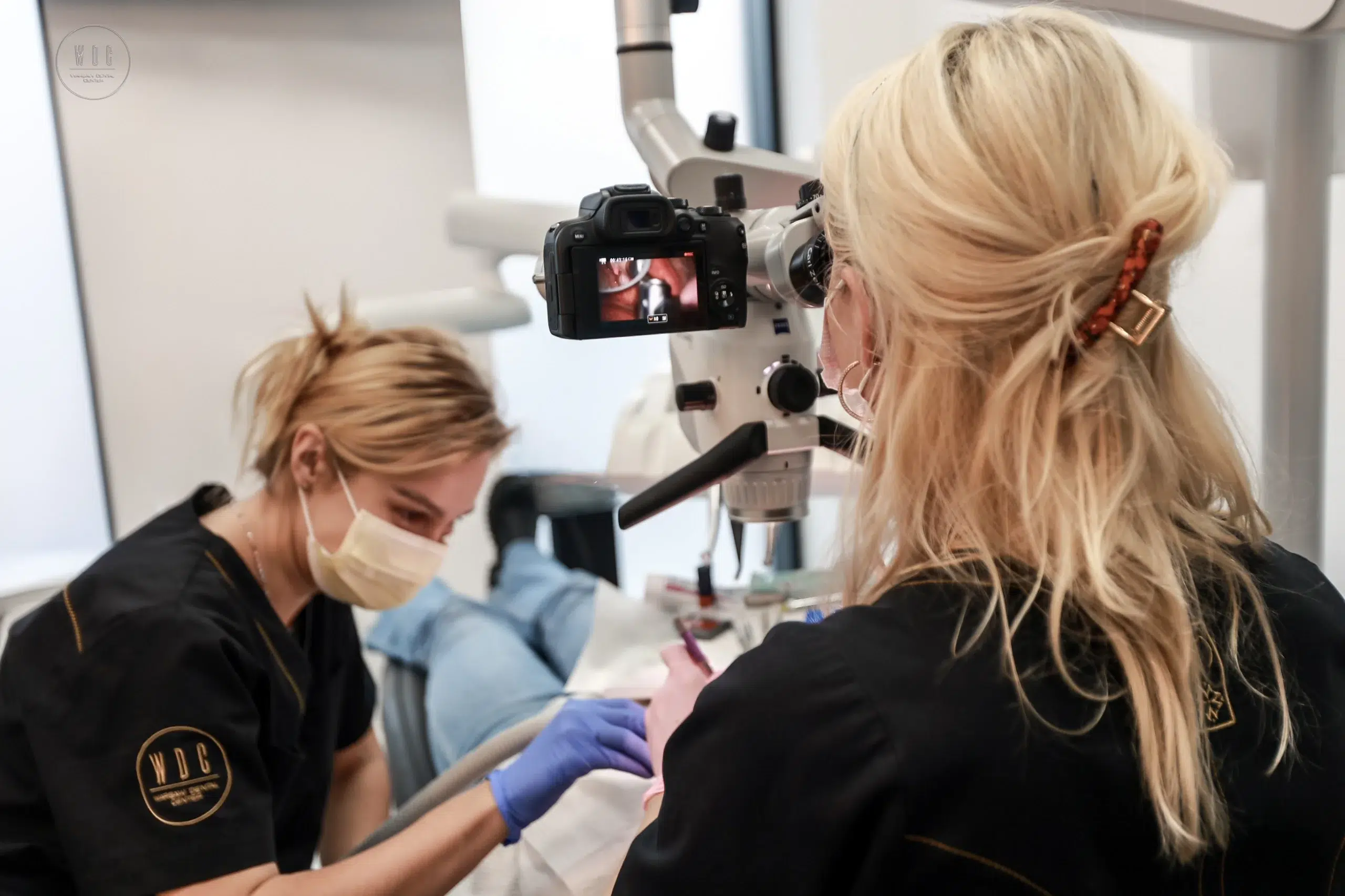
Dental microscope Modern dentistry has achieved extraordinary progress, and one of the key tools introducing a revolution in dental treatment is the dental microscope. It is not only equipment used for complex procedures, but an important element of every dental practice. Why is it worth using? A dental microscope is not only a tool with high magnification. It is a window opening the precise world of teeth, hidden details of the oral cavity, the invisibility of which to the human eye is almost inevitable. It is also a tool that gives dentists extraordinary control over every stage of the procedure, regardless of its scale. Benefits of dental treatment under a microscope: Surgical precision A dental microscope provides unrivaled precision and a detailed look at the tooth. Thanks to this, the doctor can see every detail precisely, which is unattainable in traditional examinations. Effective root canal treatment Root canal treatment, known for its complexity and demanding precision, becomes more effective with the help of a microscope. The ability to precisely view the tooth canals allows for more effective removal of any contamination, shortening treatment time and reducing the risk of complications. Diagnosis at the highest level A dental microscope enables more precise diagnosis of dental diseases. Detecting the problem at an early stage means faster action and elimination of possible ailments. Safety as a priority Dental treatment under a microscope means not only precision, but also increased safety for the patient. The microscope gives you greater control over the procedure, eliminating the risk of errors. Aesthetics and durability The use of a microscope improves the fit of the filling to the tooth, which translates into the aesthetics and durability of the final result. What does standard dental treatment look like under a microscope? It begins with a thorough examination of the patient’s oral cavity, and then the doctor uses a microscope to diagnose any pathological changes. Thanks to the use of a microscope, the procedure is more precise and less invasive for the patient, which shortens the recovery time. The dental microscope not only revolutionizes complex procedures, but also makes even routine procedures more precise and safe. It is worth trusting doctors who not only have this advanced tool, but also use it with expert hands, providing comprehensive care at the highest level.

Can migraine and toothache be related?
Did you know that there are many cases where migraine and toothache can be related? Even though these are two different ailments, sometimes they can occur simultaneously and worsen each other. Migraine is a disease that manifests itself with severe headaches, often accompanied by nausea, photophobia and/or visual disturbances. In turn, toothache may be caused by infection, damage, decay or other dental problems So how can these two different conditions be related? There are several scenarios that could explain this: Muscle tension: Some people who suffer from migraines experience muscle tension in the face and neck area, which can lead to toothache. This tension may be the body’s response to a headache. Stress and body reaction: Both migraine and toothache can be caused by stress. Stress can lead to excessive jaw tightening, which can cause toothache and at the same time make migraines worse. Dental problems and migraine: Dental infections, gum disease or even pain caused by misaligned teeth can trigger an inflammatory response in the body. This inflammatory response can, in turn, worsen the migraine or be one of its triggers. Neuralgic connections: There is a complex network of nerves in the head and neck area that allows pain from one area to radiate to another. This means that a toothache can lead to a radiating headache, and a migraine can cause a toothache. It is worth remembering that each case may be different. If you experience both a migraine and a toothache, it is worth consulting a doctor or headache specialist and a dentist. Diagnosis and treatment can be more effective when you understand whether the two problems are related or not.

Gummy Smile: Diagnosis and Treatment
Gummy smile, also known as a gummy smile, is a commonly used term a condition in which excessive exposure of the gums occurs when smiling. Inappropriate attitude between the visible red (gums) and the white area (teeth) may result in a lowered sense of aesthetics and self-confidence. Dr. Urszula Leończak is excellent at correcting this type of gummy smiles. This condition can have a variety of causes: Reasons: Tooth eruption disorder where too much gum and bone covers up teeth. Excessive tension of the upper lip muscles, which causes them to lift excessively when smiling and exposure of gums. Disturbances in the development of the jaw bones. Korekcja uśmiechu dziąsłowego może wymagać różnych podejść, w zależności od przyczyny. Leczenie ortodontyczne lub ortognatyczne może być skutecznym rozwiązaniem w przypadku krótkiej szczęki lub nieprawidłowego ustawienia zębów. Correcting a gummy smile may require different approaches, depending on the cause. Orthodontic treatment or orthognathic may be an effective solution for short or abnormal jaws tooth settings. Treatment Methods: If too much bone and gum is covering the teeth, surgery may be the best solution surgical. This may include: Gingivectomy – removal of excess gums using a laser procedure. Osteoplasty – removal of excess bones and their shaping, ensuring a lasting effect and preventing the return of a gummy smile. The above-mentioned orthodontic and surgical treatment can be supplemented with tooth reconstruction using veneers or prosthetic crowns. Thanks to this, it is possible to achieve a harmonious and natural smile, adapted to individual preferences regarding the shape and color of teeth. We invite you to the Warsaw Dental Center, where Urszula Leończak performs professional surgical procedures to correct the “gummy smile”, including both gingivectomy and osteoplasty. Our team of experienced doctors also deals with orthodontic and prosthetic solutions, providing comprehensive dental care.

The importance of dental technology in the treatment of dental caries
Nowadays, progress in the treatment and prevention of dental caries is extremely important for the health of the cavity oral. The threat posed by dental caries is being counteracted more and more effectively thanks to modern technologies in dentistry. At Warsaw Dental Center in Warsaw, we specialize in the treatment of this disease, aware of the key role that dental technology plays in combating caries. The Role of Dental Technology Dental technology is not only innovative tools, but also a modern approach to therapy dental. It covers a wide range of applications – from general care, through aesthetic and reconstructive dentistry, prosthetics and orthodontics. It is this technology that improves treatment processes and dental care, eliminating the need for outdated and time-consuming procedures. Key Aspects of Dental Technology One of the key benefits of dental technology is precise diagnostics. In During caries treatment, experts from the Warsaw Dental Center carefully diagnose the infected teeth tooth areas, which enables the effective use of appropriate therapeutic methods. Plus, thanks to these modern tools, it is possible to eliminate the causes of caries and, in unavoidable situations, painless removal of teeth affected by the disease. Reduction of Visits Thanks to Dental Technology Using advanced technological solutions, we can reduce the number of patients’ visits to the dentist, while maintaining high effectiveness of caries treatment. Thanks to dental technology in Warsaw Dental Center in Warsaw we can provide our patients with not only painless, but also effective treatment therapeutic solutions, thus supporting their oral health.

Toothache can be a sign of many diseases. What?
Do you feel a toothache but you are putting off visiting a specialist? This is a mistake! Inadequate oral care can lead to serious health problems.You will learn more about this in the “Rhythms of Health” program, where doctors from Warsaw Dental Center, including doctors Urszula Leończak, Tomasz Kupryś and Bartosz Leończak, will discuss the consequences of neglecting oral health. The program will be broadcast on January 26 at 2:55 p.m. on the TVP KOBIETA channel.
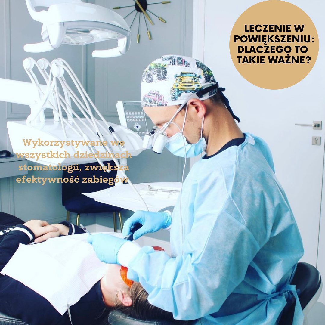
Magnification treatment: why is it so important?
Patients visiting a dentist’s office often ask themselves how it is possible that the doctor can see all the cavities or caries on the teeth and how precisely he can perform the procedures, considering that even the smallest mistake can negatively affect the results of the treatment. One of the key tools that helps eliminate the risk of complications is the use of magnification. Dentists have two main magnifying tools at their disposal: loupes and a microscope. Magnifiers are a type of glasses equipped with lenses and lighting that enlarge the image up to four times, while a microscope can enlarge the observed image up to twenty times. These tools perfectly illuminate the inside of the oral cavity, enabling each treatment to be performed with incredible precision. Thanks to them, the doctor is able to detect and treat even the smallest outbreaks of caries at an early stage of its development. When preparing teeth for crowns or veneers, minimal loss of tooth tissue is crucial – and this is where loupes play an important role. In the case of surgical procedures, they allow tooth extraction to be performed atraumatically, minimizing damage to the structures surrounding the tooth. In root canal treatment, the use of a microscope has now become a standard all over the world, because without this technology, the doctor would not be able to reach the complex internal structure of the tooth, clean it and properly fill it. The use of magnifying tools benefits not only the doctor, but also the patient at every stage of treatment, improving the quality and accuracy of procedures performed.