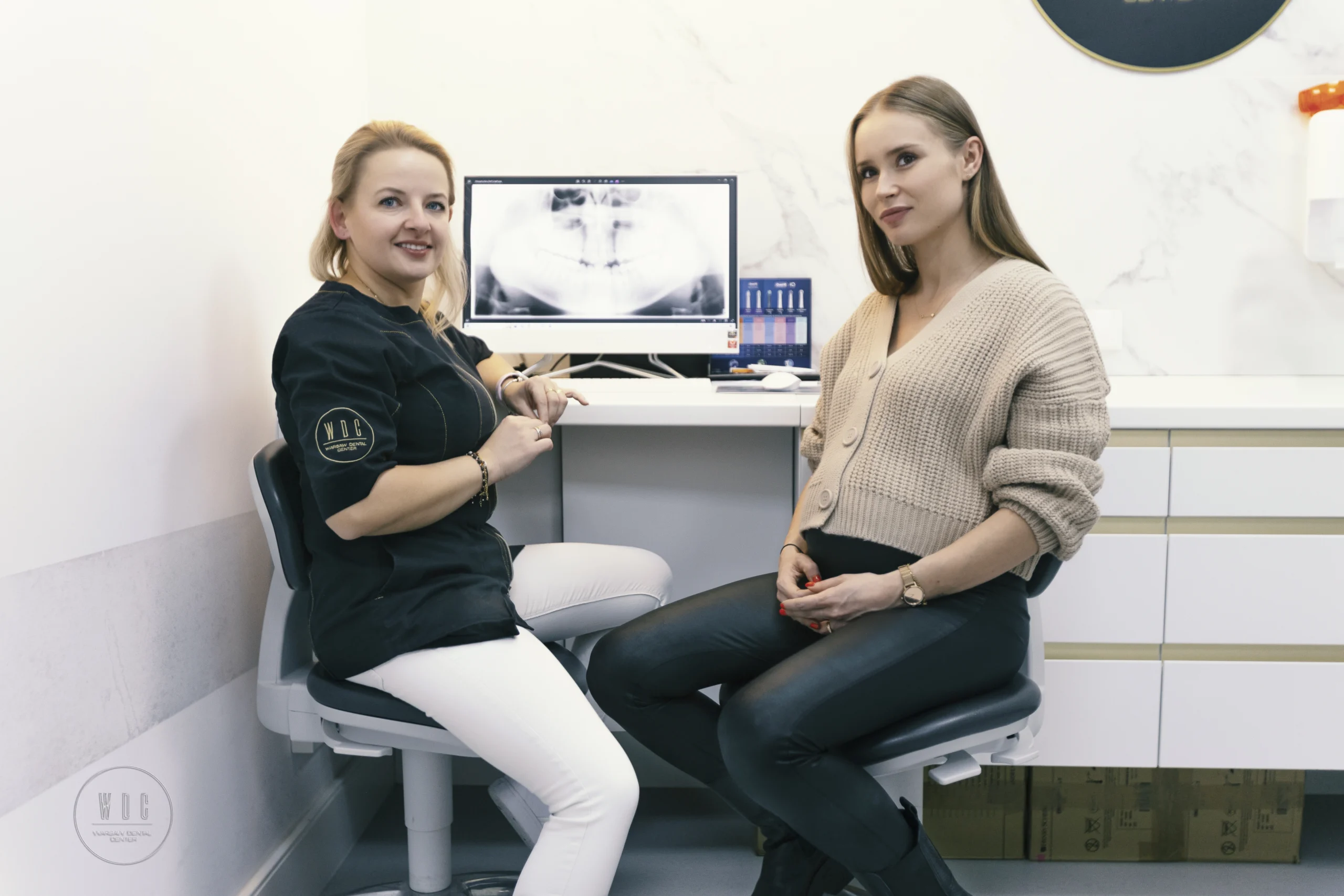
Dentist Aleksandra Kostrz discusses treatment with the patient about dental x-rays.
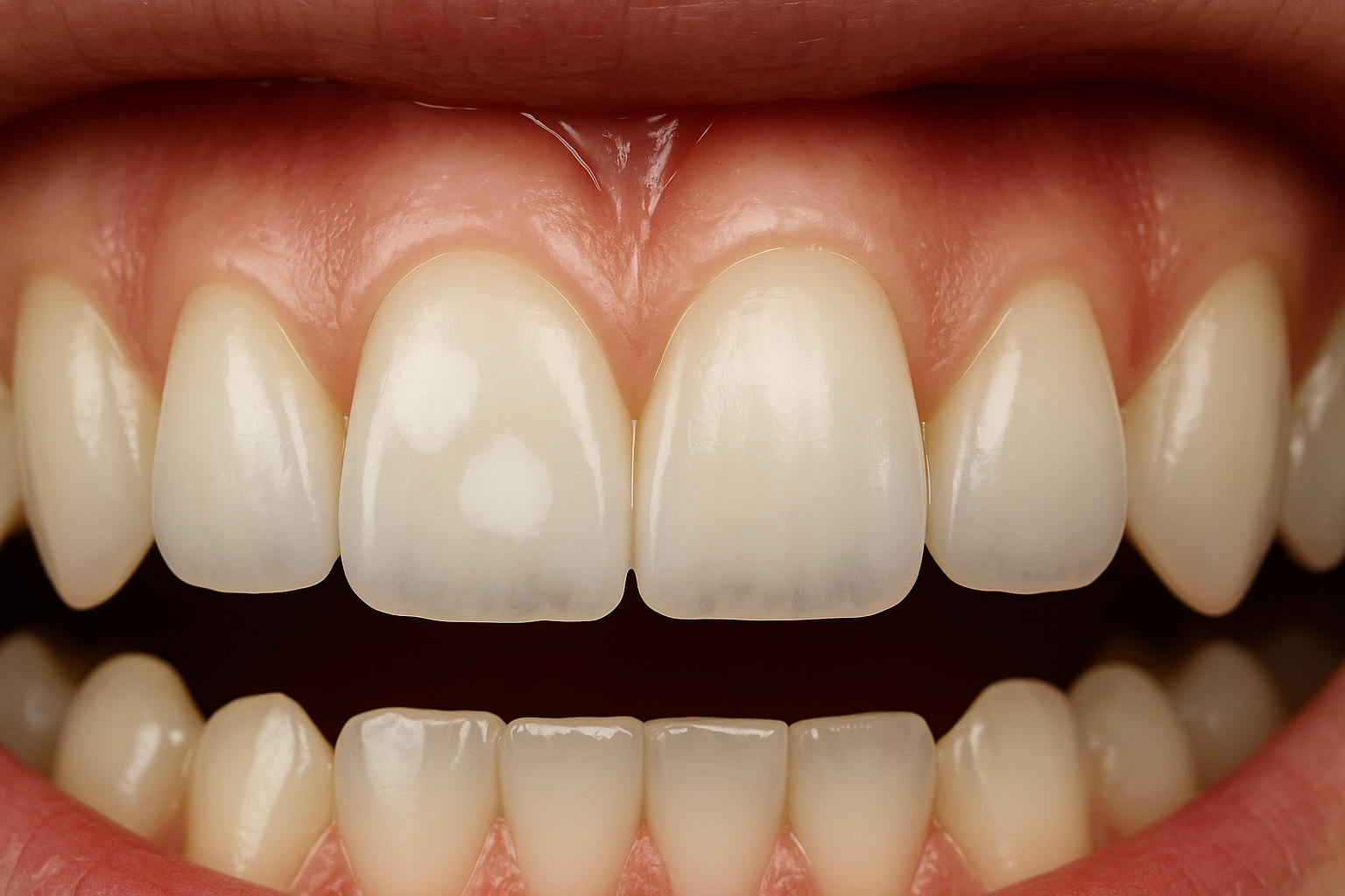
Although an experienced dentist can detect many abnormalities using his eyes, he cannot see all the details. Therefore, to obtain greater certainty, it is recommended to perform a dental pantomography. This short and effective test allows you to detect even the smallest abnormalities, including the presence of cancer.
Dental panoramic radiograph – for whom?
A panoramic radiograph is a basic diagnostic test that is an important element of the patient’s medical documentation. Its aim is to quickly assess the condition of the teeth, periodontium, jaw bones, mandible, maxillary sinuses and temporal joints. They are performed using a device known as a pantomograph.
This test detects diseases and problems invisible to the naked eye, such as:
- initial caries,
- impacted teeth, e.g. wisdom teeth,
- irregularities resulting from root canal treatment,
- cancer,
- cysts.
A dental panoramic radiograph is also extremely useful in planning prosthetic treatment, such as dentures, crowns or bridges. Before starting orthodontic treatment, it is recommended to perform a panoramic radiograph, especially if the patient has unerupted wisdom teeth, which allows for appropriate preparation for their possible surgical removal.
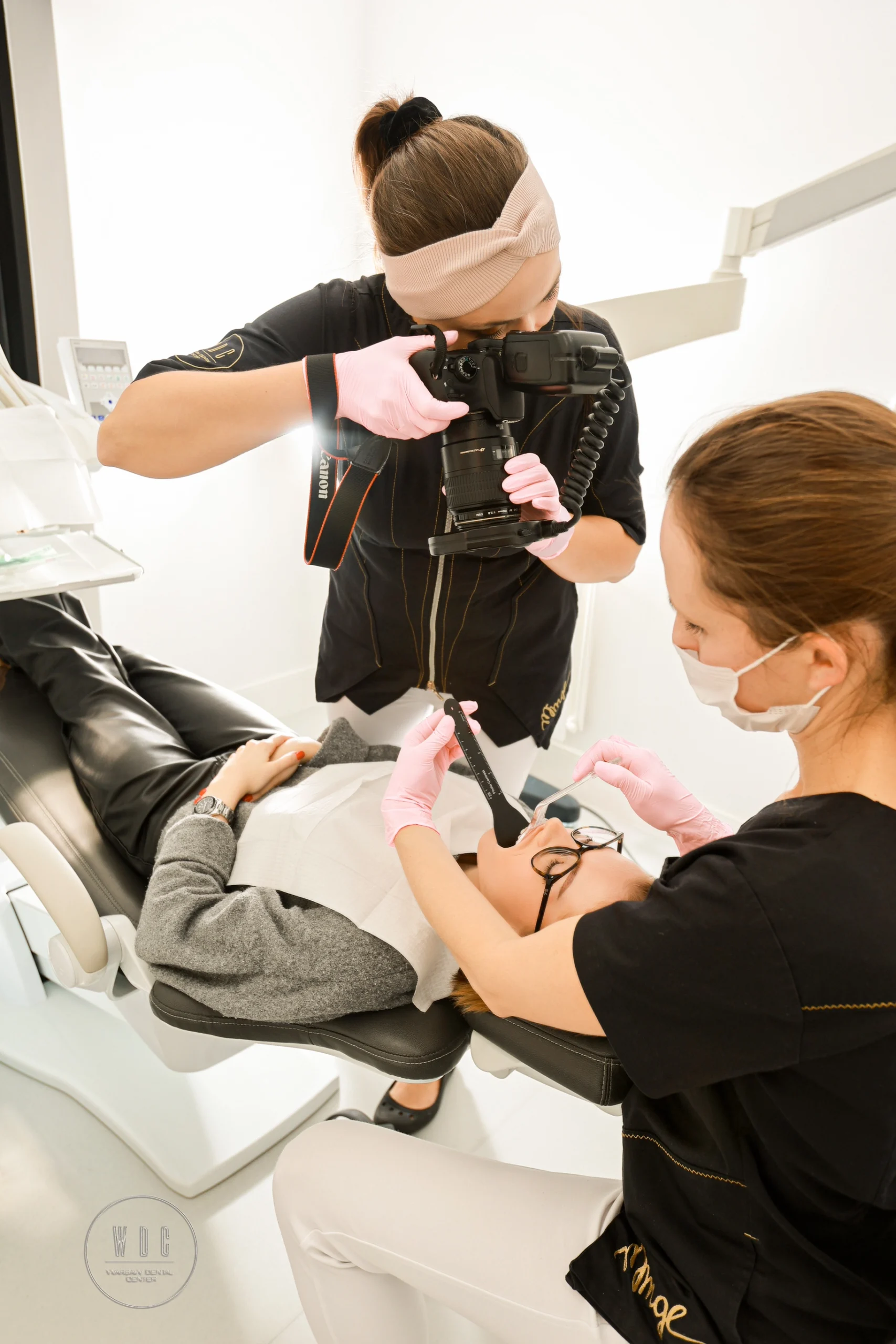
The course of the procedure of taking a panoramic X-ray
The dental pantomography procedure takes several seconds and takes place in a separate room to prevent radiation from escaping. The patient is covered with a lead blanket, which, combined with a low dose of radiation, guarantees a high level of safety. No special preparations are required, such as the need to come on an empty stomach. Only movable elements, e.g. earrings, should be removed to avoid marks on the photo. The entire procedure is painless.
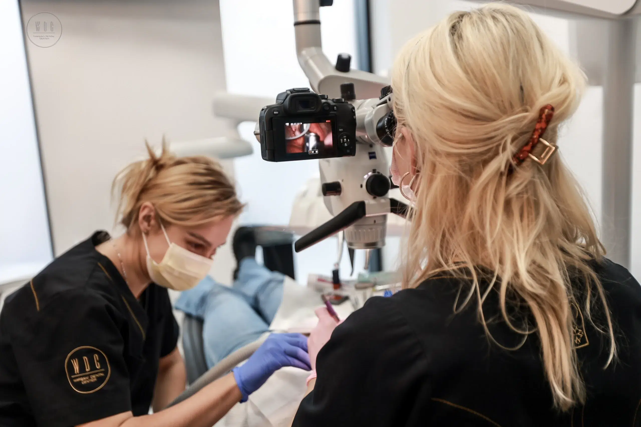
During pantomography, the radiation-generating head moves simultaneously with the recorder, which allows obtaining a precise image of a specific part of the facial skeleton. The resulting image is then enlarged by approximately 15%.