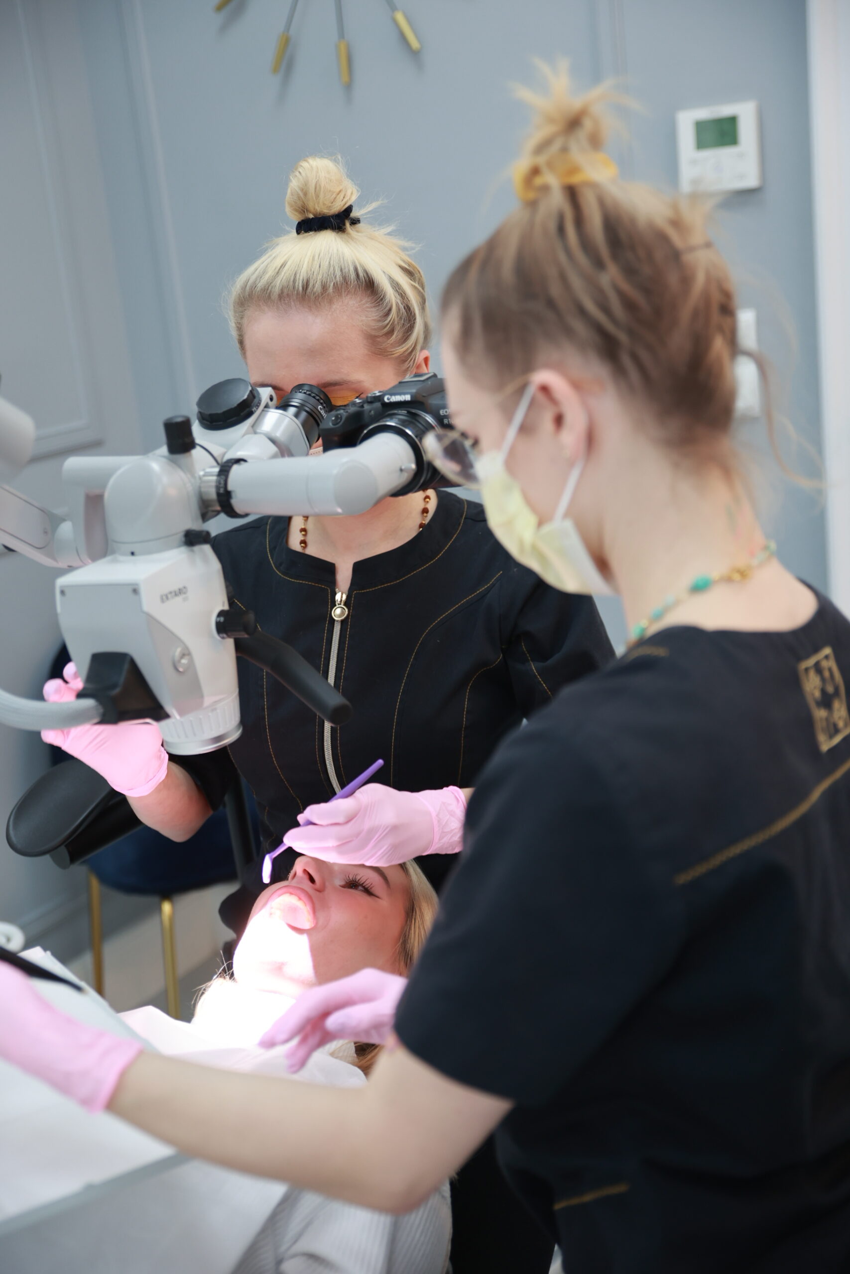
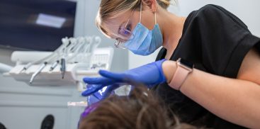
Dr. Urszula Leończak and Assistant Aleksandra Wielgat working through a microscope with a patient
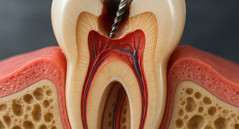
The toothache is terrible! It is a violent, throbbing pain that appears suddenly, making normal functioning impossible. This usually means that the nerve is irritated or the tooth is infected at the root. Ailments such as caries, abscesses, tooth cracks and gum problems often go hand in hand with the invasion of bacteria that damage the nerves and cause canal infections. This not only brings great pain, but can also threaten the integrity of the tooth and even lead to tooth loss. People suffering from these conditions should immediately go to a dentist who can help by performing endodontic treatment under a microscope. This advanced treatment includes a series of procedures aimed at removing all tooth content, disinfecting it and tightly securing the canals to avoid re-infection.
Microscopic revolution in endodontic treatment
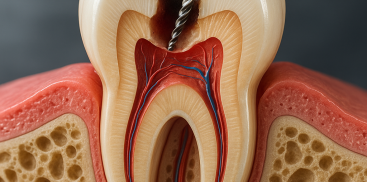
Unfortunately, even experienced dentists are not always able to see exactly what is happening in the tooth canal, especially due to the individual shape of each tooth. However, there is innovative endodontic technology – treatments performed under a microscope. At Warsaw Dental Center, this microscopic equipment has revolutionized endodontic treatment, opening up new possibilities for diagnosis and treatment, enabling the solution of difficult cases.
Using the latest technologies and an interdisciplinary approach, Warsaw Dental Center develops personalized methods of endodontic treatment under a microscope. These can be simple root canal treatments, single-root treatments or multi-root treatments that restore or maintain the internal functionality of teeth in the long term by removing the nerves or dental pulp. The presence of an endodontic microscope in our clinic allows us to achieve the highest quality of dental treatment.
Precision and effectiveness thanks to a microscope
Root canal treatment under a microscope, performed by a professional endodontist, can usually be completed in one visit. The microscope provides direct visibility of all details and pathogens, which translates into excellent results. The images are extremely clear and can be enlarged up to 30 times, and strong lighting allows you to see even the smallest details invisible to the naked eye. This is extremely important for correct diagnosis and treatment. The light is directed exactly where the doctor is working, no shadows are created, and the colors are reproduced faithfully.
Benefits of using an endodontic microscope
- Detection of unusual root canals.
- Locating perforations in the tooth root and sealing them.
- Identification of microcracks and fractures along the length of the tooth.
- Detection of root canal orifices.
- Identifying and treating canals that are difficult to access (calcified).
- Repair of incorrect root canal treatments.
- Observing cracks in the root canal, simplifying and removing them.
- Saving teeth that would otherwise have to be extracted.
- Creating photos or videos during endodontic treatment, which facilitates communication with the patient.
The opinions of dentists using this microscopic technique are extremely positive. They emphasize that they are less tired both mentally and physically. Patients feel comfortable and safe, and the effects of treatment are quick and lasting. Thanks to the endodontic microscope, endodontic treatment becomes more precise and effective, which translates into the health and comfort of patients.
